
Welcome to this week’s Research Roundup. These Friday posts aim to inform our readers about the many stories that relate to animal research each week. Do you have an animal research story we should include in next week’s Research Roundup? You can send it to us via our Facebook page or through the contact form on the website.
- Wound glue could replace internal sutures. Traditional sutures, staples, and wires are difficult to properly place in some areas of the body and do not close wounds immediately, which increases the risk of infection. Researchers have developed a human protein-based hydrogel sealant called MeTro that seals wounds in 60 seconds. Unlike other currently available sealants, MeTro contains all the characteristics of a successful surgical glue: it is elastic, adhesive, non-toxic, and biocompatible. It has successfully healed incisions in the arteries and lungs of rats and wounds in the lungs of pigs. MeTro remains stable while the wound heals, even when applied to tissues that constantly expand and relax, and then degrades without any traces of toxicity. Published in Science Translational Medicine.
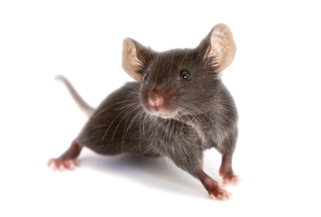
- Vaccine for virus induced Type 1 diabetes developed. The incidence of Type 1 diabetes is increasing while the etiology of the disease itself is unknown. One implicated environmental cause is enterovirus and in particular Coxsackievirus B (CVB). To investigate this link further, researchers inoculated mice with a CVB1 vaccine and tested whether this prevented the development of diabetes in a clinically relevant model. Prof. Malin Flodström-Tullberg stated: “these exciting results showing that the vaccine completely protects against virus-induced diabetes indicate the potential that such a vaccine has for elucidating the role of enteroviruses in human Type 1 diabetes.” This research was published in the journal Diabetologia.
- Genetic component of a rare form of liver cancer confirmed. Fibrolamellar hepatocellular carcinoma (FL-HCC) is a rare liver cancer, with an incidence rate of rate of 1 in 5,000,000. It usually occurs in adolescents and young adults who have no history of liver disease. The cause of this disease has been identified as a chimeric gene, where the two genes Dnajb1 and Prkaca fuse together. In the present study, these researchers used the CRISPR gene editing technique to create a mouse model with this chimeric gene. They found that these mice develop liver tumors similar to humans and that this chimeric gene was by itself sufficient to cause FL-HCC cancer. This research was published in the journal Proceedings of the National Academy of Sciences.
- Role of muscle in tissue regeneration. “One of the central mysteries in organ and tissue regeneration is, how do animals initiate all of the cellular and molecular steps that lead to regeneration?” states Peter Reddien. To investigate this question, these researchers turned to the planarian Schmidtea mediterranea. myoD. They found that longitudinal muscle fibers are responsible for initiating tissue regeneration while circular fibers aid with the formation of the correct pattern. These results underscore the relatively ignored role of different muscle fibers in tissue regeneration. This research was published in the journal Nature.
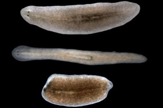
Credit: Lucila Scimone/Whitehead Institute
- Refinement of stroke induction method in mice promises a reduction in the number of animals used. Most strokes are caused by an abrupt blockage of arteries leading to the brain — ischaemic stroke. To model this in mice, the middle cerebral artery (MCA) of the brain is blocked. The limitation of this approach is that it often results in inconsistent lesion sizes – leading to a large number of animals being needed in order treatment effects being observed. In order to reduce variation in lesion size, these researchers modified the existing protocol so that incisions made in the blood vessels are repaired with tissue pads instead of these vessels being permanently tied off. This resulted in smaller variation in lesion size – and ultimately smaller numbers of animals being needed to detect statistically significant results. This study was published in the journal Disease Models & Mechanisms.
