
The 2016 Lasker Awards have highlighted some great discoveries and the scientists behind them. This guest post by Samuel Henager, a graduate student at Johns Hopkins University, investigates how animal studies contributed to the discoveries celebrated by this years’ Lasker Awards.
Basic Medical Research Award
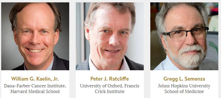
The 2016 Albert Lasker Basic Medical Research Award was awarded to William G. Kaelin, Jr. of Dana-Farber Cancer Institute, Harvard Medical School, Peter J. Ratcliffe of University of Oxford, Francis Crick Institute, and Gregg L. Semenza of Johns Hopkins University School of Medicine for their work in discovering how cells sense and respond to changes in oxygen levels.
Oxygen is crucial for survival, but at the same time, too much can be toxic for cells and damage DNA and proteins. Thus, it is crucial for cells to be able to sense and respond to the concentration of oxygen in its environment. Semenza and Ratcliffe discovered that under low-oxygen conditions the protein hypoxia-inducible factor-1a (HIF-1α) turns on many genes. Subsequently Kaelin and Ratcliffe discovered that under high-oxygen conditions, an enzyme called prolyl hydroxylase caused HIF-1a to be destroyed by the protein von Hippel-Lindau (VHL). VHL is mutated in von Hippel-Lindau disease, which is characterized by large tumors made of blood vessels. In the disease, HIF-1α levels are artificially high due to a defective VHL protein, thus tricking the body into thinking it needs more oxygen, and mistakenly growing unneeded blood vessels to carry oxygen to seemingly low-oxygen tissues.
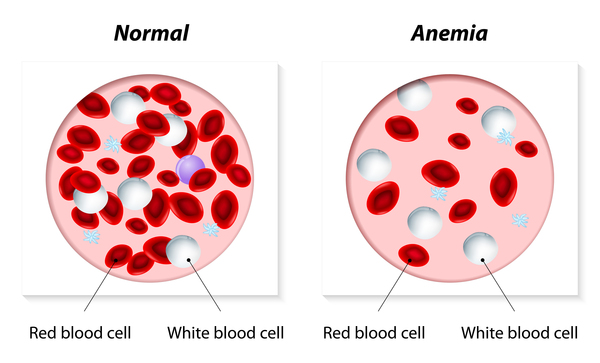
The discovery of the full pathway for how cells respond to differing levels of oxygen has fueled ongoing research. Stopping the destruction of HIF-1α can help with anemia, a condition where low iron makes red blood cells less effective at carrying oxygen, by increasing the production of red blood cells. There are also cancer treatment applications, as some tumors’ survival depends on HIF-1α to spur the development of new blood vessels.
HIF-1α is conserved across a wide variety of species, and many animal models played a crucial role in the discovery of HIF-1α and its function. The first study by Ratcliffe that indicated a wide-spread response to low oxygen used multiple cell culture systems from monkey, pig, Chinese hamster, rat, and mouse cells. In later studies by Kaelin, Ratcliffe, and Semenza, reticulocytes—precursors to red blood cells—from rabbits were used to generate HIF-1α protein to study in vitro. Xenopus laevis (frog) cells were used to study how prolyl hydroxylase was involved in the destruction of HIF-1α. C. elegans (roundworm) were used to investigate how mutations in VHL affected a whole organism’s ability to respond to low oxygen levels. Mice were used to study how HIF-1a might be involved in anemia. The discoveries celebrated by this award have fueled new avenues of research and the development of novel therapies, and animal models will surely continue to be a key part of this story.
Clinical Medical Research Award
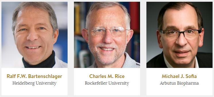
The 2016 Lasker-DeBakey Clinical Medical Research Award was given to Ralf Bartenschlager of Heidelberg University, Charles M. Rice of Rockefeller University, and Michael J. Sofia of Arbutus Biopharma for their work in developing a system to replicate Hepatitis C virus (HCV) in the lab and for using this system to develop new drugs to cure Hepatitis C infections.
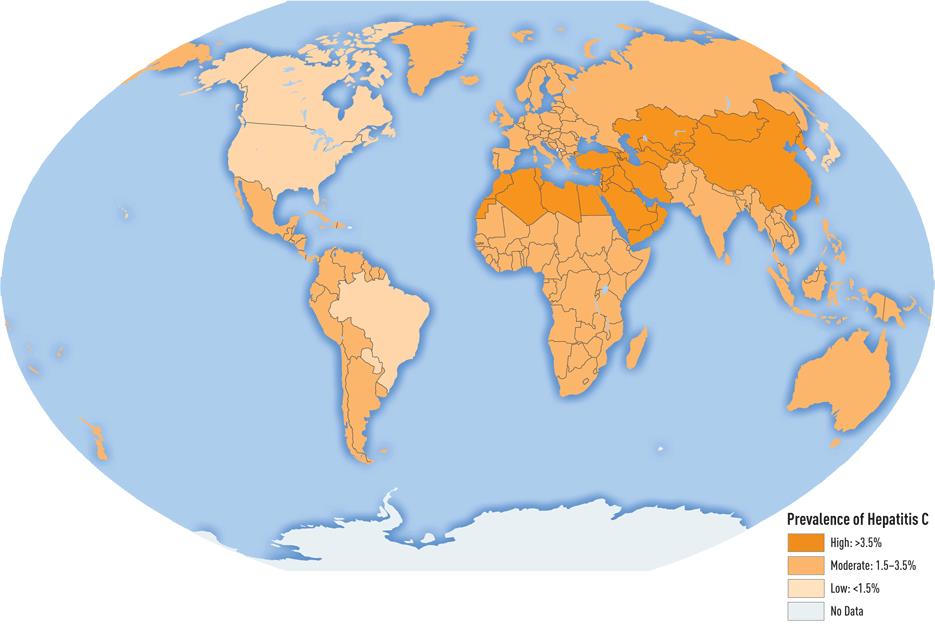
Hepatitis C can be a devastating illness, leading to cirrhosis of the liver, liver failure, and liver cancer. Previous treatments to fight the infection were highly toxic and did not effectively cure the person from disease. Drs. Bartenschlager, Rice, and Sofia all contributed to discovering a much safer, effective treatment for Hepatitis C.
The virus responsible for Hepatitis C was identified in 1989. For many years after its discovery, scientists struggled to create a strain of HCV that could replicate under laboratory conditions so that they could study the components and life-cycle of the virus in order to develop treatments or a vaccine. In the late 1990s, Dr. Rice recreated the full genetic sequence of the virus, and used this sequence to infect chimpanzees with the virus. At the time, chimpanzees were the only animal model for hepatitis, and he needed to make sure that the sequence he had identified was capable of replicating and causing disease. At the same time, Dr. Bartenschlager was attempting to infect liver cells using the newly identified sequence, but never detected replication. He was unsuccessful until he inserted a drug-resistance gene into the virus which allowed infected cells to survive when the culture was treated with a lethal drug. He also identified several mutations in the virus that allowed for better replication. With this improved sequence he was able to successfully infect a liver cell line with hepatitis C, which allowed scientists to study the virus in depth and begin to develop therapies for the disease. Dr. Sofia led a team of pharmaceutical researchers that developed a novel therapy for hepatitis. This new therapy is able to cure chronic hepatitis for many patients, who otherwise would be at risk for liver failure and liver cancer.
This is not only a great story of finding a cure for what can be a devastating disease, but also a great example of the value of non-human primate (NHP) research. The cellular replication system developed by Dr. Bartenschlager was important for developing drugs and studying the life-cycle of the hepatitis viruses, but for many years, the only way to study HCV was in a chimpanzee model. Chronic hepatitis C infection can lead to liver cancer, but how the virus or disease contributes to cancer development is not known. Humanized mouse models of hepatitis have been introduced in recent years, and scientists continue to work to improve their accuracy. These mouse models will be crucial as scientists work to unravel the remaining questions surrounding this disease, and work to develop effective treatments and vaccines.
Samuel Henager
Graduate student, Johns Hopkins University
