
Welcome to this week’s Research Roundup. These Friday posts aim to inform our readers about the many stories that relate to animal research each week. Do you have an animal research story we should include in next week’s Research Roundup? You can send it to us via our Facebook page or through the contact form on the website.
- 3D printed ovaries produce healthy offspring in mice. In a proof of principle experiment, scientists have succeeded in removing and replacing a mouse’s ovary with a bioprosthetic replacement. These 3D printed ovaries housed immature mouse eggs, permitting the mice to ovulate and to also give birth to healthy pups. These mice were also able to successfully nurse their pups.The 3D printed ovaries were made of gelatin and is safe to use in humans. This research is highly applicable to “women who have undergone adult cancer treatments or those who survived childhood cancer and now have increased risks of infertility and hormone-based developmental issues.” This paper was published in Nature communications.
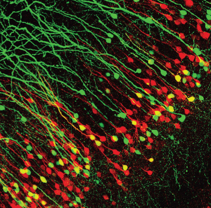
- New study investigating how the brain encodes fear memory could lead to novel therapeutics aimed at reducing pathological fear in PTSD. Using mice, a tracing method which highlights neurons in the brain in different colors, electrophysical and optogenetic methods — these researchers that as much as 17% of neurons projecting to the hippocampus also projected to the amygdala and the mPFC. The authors explained “that the acquisition (encoding) and retrieval of contextual fear memory requires coordinated neural activity in the hippocampus, amygdala and mPFC. The hippocampus encodes context cues, the amygdala stores associations between a context and an aversive event, and the mPFC signals whether a defensive response is appropriate in the present context.” This study was published in the Journal of Neuroscience.
- A simple blood test detects radiation exposure in nonhuman primates. A report in the Science Translational Medicine describes how levels of three microRNAs (miRNAs, which are small non-coding RNA molecules found in plants and animals that regulate gene expression) can be detected in blood and other bodily fluids with 100% accuracy. Two other miRNAs can predict whether the radiation exposure will be fatal. This new development could help triage victims of nuclear disasters, like the one in Fukushima, Japan, in 2011. The assay could be deployed in the field with limited expertise or equipment.
- Large families and strong social ties help animals live longer. A large study of a naturalistic population of rhesus monkeys on an island off of Puerto Rico shows that adult monkeys with more relatives in their social network have a better life expectancy. Rhesus monkeys live in matriarchal societies, meaning that families are formed around adult females and their offspring and other relatives. By examining demographic data on over 900 monkeys across a 21 year period, researchers found that each extra female relative reduced a prime-aged female macaque’s chances of dying in one year by 2.3%. Because humans and rhesus monkeys shared a common ancestor about 25 million years ago, “we can take clues from these distant cousins about how humans might have existed in pre-industrial societies,” said Dr. Laurent Brent, Leverhulme Early Career Fellow and Lecturer at the University of Exeter and lead author on the study. “Human societies are hugely complex, and factors such as culture and access to healthcare make it hard to study the impact of a single factor like social relationships on survival.” The study was published on Tuesday in Proceedings of the Royal Society B.

- Breakthrough findings in bone marrow transplants, melanoma and anemia thanks to zebrafish. Zebrafish are important models for studying human disease. Dr. Leonard Zon, a researcher at Harvard University, is studying blood diseases and cancer. The embryos are transparent so that organ development can be visualized as it happens. Another advantage is that the mothers have many eggs allowing the study of the genetics of blood diseases and cancer that are passed on within families. This provides the ability to find similar genes in humans that cause those diseases. His work has led to two drugs now treating patients. One increases stem cells to help bone marrow transplant patients and the other treats patients with metastatic melanoma. A third drug is now in clinical trials with hopes to help people with a very rare type of anemia.
- A new link in the gut-brain axis has been identified. A study in mice found that bacteria in the gut are implicated in cerebral cavernous malformations (CCMs), brain defects that occur in as many as one in 100 people. These malformations are blood-filled bubbles that protrude from veins in the brain and can leak or burst at any time. The researchers discovered that Gram-negative bacteria, which are typically found in the gut, accelerate CCM formation, particularly in animals that increase the expression of a certain gene, toll-like receptor 4 (or TLR4). A single course of antibiotics permanently altered CCM susceptibility in mice. This research identified unexpected roles for the microbiome, and is among the first to provide convincing evidence that these bacteria may initiate diseases in seemingly unrelated organs. The study was published on Wednesday in Nature.
- After decades of research, lab-grown blood stem cells have been produced providing “hope to people with leukaemia and other blood disorders who need bone-marrow transplants but can’t find a compatible donor.” Using standard methods, these researchers transformed cells into induced iPS cells. They next inserted seven transcription factors into the genome of of the iPS cells. Injecting these modified human cells into mice. Twelve weeks later these cells had transformed into “progenitor cells capable of making the range of cells found in human blood, including immune cells.” This research was published in the journal Nature.
- A new gene therapy method for bone regeneration has been developed to serve as an alternative to bone grafts. Currently bone graft technology leaves many patients with fracture nonunions; where fractures are repaired but fail to fully heal. Furthermore, patients are often lead to long-term hospitalization and repeated surgeries. However, a promising gene therapy solution has been developed in mini-pigs, a clinically relevant large animal model. Researchers implanted a collagen scaffold into a critical-sized bone fracture of mini-pigs to recruit endogenous bone stem cells. Two weeks later bone morphogenetic protein-6 (BMP-6) DNA and microbubbles were injected into the fractures. This lead to bone regeneration and fracture healing over 6 weeks. This minimally invasive method shows great promise for human translation. This study was published in the Journal Science Translational Medicine
Gene therapy for bone regeneration. Source: M. Bez et al., Science Translational Medicine (2017)
