
April 5th, 2021
Professor Christopher Petkov
There is a misconception often propagated by individuals or organizations that do not see much benefit to animal research. They would have the public believe that scientists shock an animal’s brain for little to no good reason. This claim is not true and we encourage you to call it out with #FactCheckNeeded @SpeakofResearch whenever you see it.
Here, we fact check the claim and consider:
- How and why scientists and clinicians electrically stimulate the brain
- Why it remains important to study the impact of stimulating the brain with nonhuman animals (illustrated with a few recent peer-reviewed studies)
Brain neurons communicate using electro-chemical potentials
Stimulating the brain electrically makes sense because brain neurons communicate through electricity and chemical molecules. The electrical potentials of neurons can also be recorded, which allows us to ‘listen’ to how the brain works.
Areas of the brain are connected via networks of neurons, much like networks of electrical wires carry electricity to houses. Neurons communicate using, in part, chemical molecules called neurotransmitters. Neurotransmitters released by neurons bind to receptors present on the body of receiving neurons, their cell membranes. Because neurons are negatively charged on the inside, when a neurotransmitter binds to certain types of receptors, this opens up the receptor channels and can then let positively charged particles into the neuron. When enough positively charged particles (like sodium: Na+) enter the neuron, they change the neuron’s electrical potential, depolarizing it. This leads to an electrical action potential that quickly propagates down a neuron towards other neurons, resulting in the further release of neurotransmitters, thereby continuing the electro-chemical communication between connected neurons.
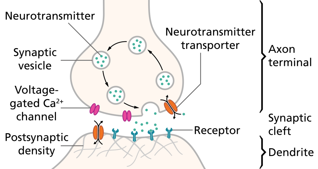
Practically, this means that scientists can do two things. They can use electrodes to record the electrical potentials of neurons, if for example they need to know what a neuron is responding to. Scientists can also pass a small current through the electrode to stimulate the neurons, in order to initiate their communication with other neurons in the brain to which they are connected. This often requires animal research because direct neuronal recordings in humans are rare and can only be conducted as part of clinical treatment for neurosurgery patients.
If we were recording from inside of a neuron, we would see that it elicits a very small potential in the millivolt range (thousandth of a Volt). These potentials when generated by many thousands of neurons can also be measured at the surface of the head using ElectroEncephalography (EEG, which records the brain’s electrical potentials) or MagnetoEncephalography (MEG, which records the change in the magnetic field perpendicular to the electrical current flow in the brain).
Stimulating neurons can be done by microelectrodes, which are typically smaller than a fraction of a millimeter. Or it can be done with electro-magnetic coils at the surface of the head using, for example, Transcranial Magnetic Stimulation (TMS) or Transcranial Direct Current Stimulation (tDCS).
Brain stimulation is an important treatment for neurological and psychiatric disorders
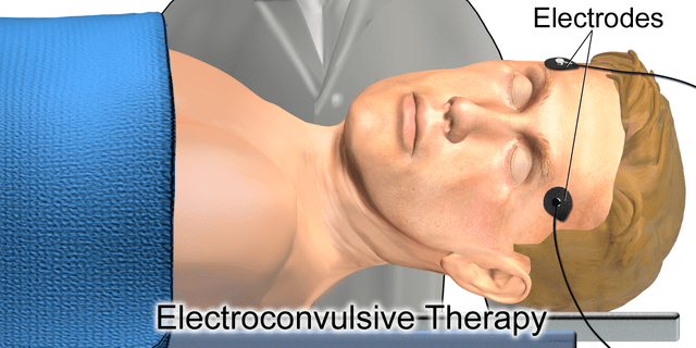
One of the strongest and most effective forms of electrical brain stimulation is called ElectroConvulsive Therapy (ECT). ECT is conducted only in anesthetized (unconscious) patients and to treat debilitating and life-threatening mental health conditions like drug-resistant clinical depression. There are also many myths and misconceptions about ECT. Dr. Kala Bailey at UT Southwestern Medical Center provides more information on ECT and debunks common myths associated with it in this article.
Current research on ECT is exploring how to best apply it to make it more effective and reduce its potential side effects. This is being done in combination with computer modelling and brain imaging to both predict and evaluate how to make ECT more effective for individual patients. Animal research provides an important complement for understanding ECT: How does it work? How can we improve it? And how can it be implemented safely in humans? Magnetic Seizure Therapy (MST) is being investigated as an alternative, but still remains less effective than ECT in severe drug resistant individuals. Thus even MST could benefit from further research understanding how it works and how to improve the patient’s treatment response.
Other forms of electrical brain stimulation may require a surgical procedure to implant microwires to electrically record from or stimulate specific parts of the brain. For instance, in drug-resistant epilepsy a patient’s brain is clinically implanted with recording electrodes to localize the seizure inducing sites, which once identified would then be surgically removed (resected).
A patient with implanted electrodes would typically be monitored for a couple of weeks as clinicians obtain enough information to guide their decisions about which brain area(s) need to be resected in order to treat the patient’s epilepsy. During this monitoring period, clinical and research teams may stimulate different parts of the brain to try to confirm that these regions can initiate seizures or to understand which brain regions to avoid in order not to leave the person with cognitive problems after the surgical treatment.
Here is a video from a scientific study conducted with an epilepsy patient by a Stanford University research team. It shows the experience of the epilepsy patient during the monitoring period, including how stimulation of a contact over visual cortex strikingly changes the patient’s ability to recognize a face.
Clinicians that neurosurgically treat epilepsy must carefully balance between an effective surgical treatment and preserving the patient’s language, cognitive or other abilities (such as recognizing faces, as seen in the Stanford patient above). If the epilepsy treatment goes well, the patient may then become seizure free (or nearly so) with as few cognitive side effects as possible, being able to work, drive and lead a more normal life.
Other forms of electrical stimulation for treatment include cochlear implants, which are electrodes implanted in the damaged cochlea within the ear. When the cochlear implant is stimulated this can help neurons in the intact auditory system to detect sounds, helping patients with cochlear implants to hear sound. Deep Brain Stimulation (DBS) is another form of brain treatment. Its history and reliance on animal research, including in nonhuman primates, have been previously described in articles by Speaking of Research here and here. DBS requires an implanted electrode, and it is used to treat brain disorders such as Parkinson’s disease. DBS implantation is usually performed in awake patients to help the surgeon guide the insertion location and the efficacy of the treatment.
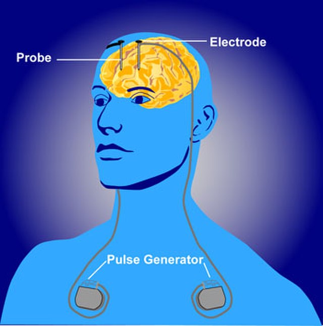
Visualization of Deep Brain Stimulation
Why is it important to stimulate the brains of nonhuman animals?
Research with nonhuman animals, including monkeys, has been and remains instrumental in a number of ways as can be read here. First, animal research is required to lay the foundation for any future treatment. This means that there may be basic or fundamental science information that needs to be obtained first, even if this does not have an immediate impact on a potential treatment, as most such research would not yet would still be useful. Second, animal research can generate scientific knowledge on how generalized or localized the brain stimulation is and how it works, including what impact it has on the intended brain pathways and neuronal networks. Third, the animal research can also explore the safe use of the technology in humans or how to improve stimulation even after the technique has started to translate to greater use in humans.
A powerful combination: Combining stimulation with brain imaging
Brain electrical stimulation is now more readily being combined with brain imaging. A brain imaging system like that using functional Magnetic Resonance Imaging (fMRI) can show which brain areas use more oxygen when a specific part of the brain is stimulated.
An analogy here is sending signals down the brain’s neuronal highways and using brain imaging to visualize how the electrical stimulation affects other parts of the brain along those highways.
Here is an image from a recent comprehensive review on nonhuman primate brain stimulation approaches, how they are combined with brain imaging and the potential usefulness for humans.
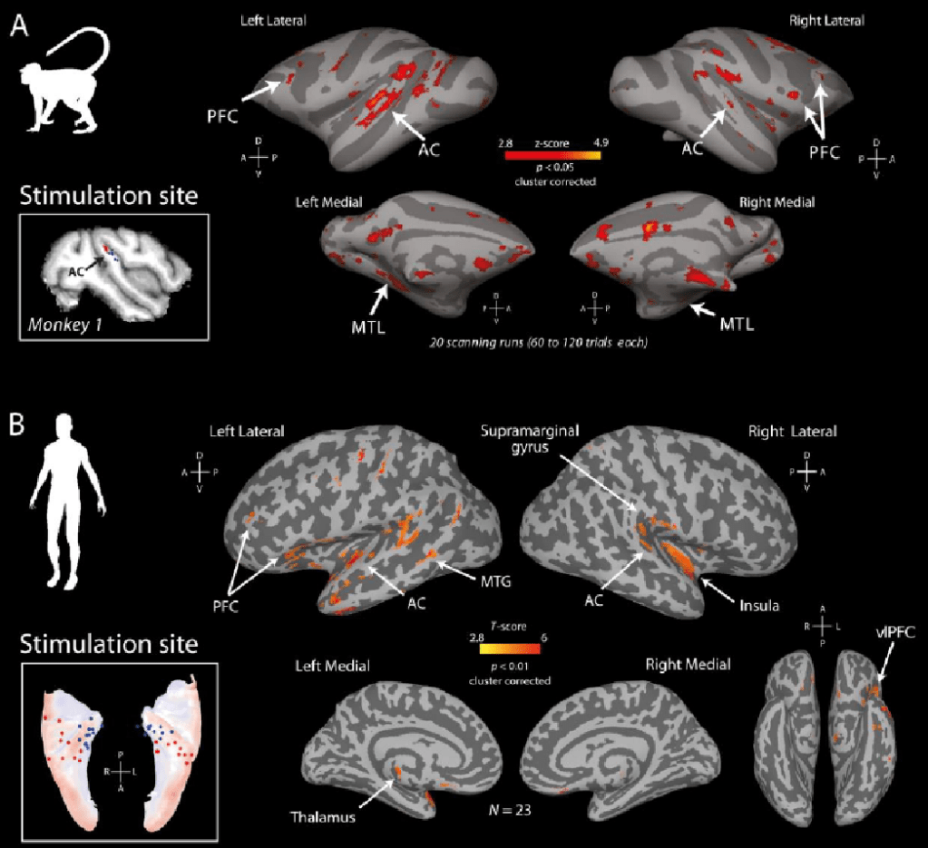
How brain imaging allows visualizing stimulation effects. (A) shows brain imaging activity in red resulting from stimulating the auditory cortex in monkeys. (B) shows the same but in human neurosurgery patients during stimulation of electrical contacts in the human auditory cortex.
Why is it important to stimulate the human and monkey brain and compare effects
Speech and language are unique to humans, thus an important scientific challenge, if animal research is to be as informative as possible for the human condition, is understanding the extent to which the human brain specialized for what we are doing now: communicating via language. Our language abilities also help us to better remember. For instance hearing someone say that “the singer sounded just like a nightingale” not only helps us to better remember the qualities of the singer but also to imagine what their singing is like even if we have never heard it.
A recent study in humans and monkeys directly compared the effects of brain stimulation of areas that in humans are involved in language and memory with parallel study of homologous areas in the monkey brain (see Figure above; Video Abstract to the paper). They then compared the results obtained in humans to those in the monkeys. In so doing, the neuroscientific and neurosurgery teams found that the brain’s pathways for language and memory are remarkably similarly wired in both humans and monkeys, which implies that both species’ brains use an evolutionarily conserved brain highway system that likely dates to a common ancestor that they shared over 25 million years.

Male rhesus macaque. Source: Kathy West.
In other words, the brain highways even for language and memory in humans are based on a deeply conserved brain system that is also present in monkeys. This means that monkeys are an important neurobiological model, even for aspects of the human language and memory system that a priori we may have thought specialized too much in humans to find a realistic animal model.
The authors found much more subtle, potentially human-unique effects, with some of the human brain stimulation activity being stronger in one brain hemisphere than the other. By comparison, the effects in the monkeys were largely symmetrical (mirrored) across the two hemispheres.
The study also generated unique new brain scanning data and information that was shared across the globe as part of transparency and open data sharing initiatives like the PRIMatE Data Exchange (PRIME-DE). Data sharing efforts such as these aim to support and inspire further discovery with the shared data by the international scientific community and help to ensure the best use of animal research to inform neuroimaging work with humans, for example.
Brain stimulation and animal research remain indispensable
I conclude with a few examples of continuing scientific developments and why they require animal research. Safety issues that have plagued the ability to conduct brain stimulation and neuroimaging in humans are now being better addressed, in large part because of the bedrock of scientific information available in nonhuman primates and rodents. This work is ongoing and will continue to depend on animal research.
There are also now unique brain stimulation and imaging resources available in humans, so that scientists, or those of you interested in seeing brain imaging results, can visualize the effects of stimulating brain areas such as the amygdala (which is implicated in emotions and affect), the hippocampus or auditory cortex. Parallel work in monkeys continues to be needed because it provides an important bridge between the human studies and information that cannot be conducted in humans and thus requires research with nonhuman animals.
Shocking news or #FactCheckNeeded?
In conclusion, the notion propagated by some individuals or organizations that scientists shock animal brains for little to no good reason could not be further from the truth. That claim itself seems nothing merely than an attempt to “shock” the public in order to disseminate misinformation to put scientists and their work in an unfavorable light. When you see it please call it out with #FactCheckNeeded @SpeakofResearch. We also welcome you to do some fact checking as a guest writer for Speaking of Research.
